Co-design and implementation of ultrasound transducers and electronics allows one to integrate front end system and signal processing capabilities to enhance the performance of the overall imaging system, enable new imaging tools, as well as reducing the size and cost of existing systems. We have been working on several applications of these integrated microsystems where we combine our knowledge of transducers, integrated circuits and image/signal processing.
Wireless Intracranial Ultrasound (WICUS)
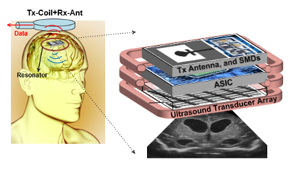
The aim in this project is to provide on-demand ultrasound imaging of the brain to post-operatively monitor treatment efficacy of brain tumors over time. The approach is to use a wirelessly powered implant placed inside the skull during the operation which can provide images, intracranial pressure, and blood flow information wirelessly transmitting the data to the outside system. This enables use of higher frequency ultrasound for higher image resolution while avoiding the poor penetration of ultrasonic waves through the skull.
Intravascular Ultrasound (IVUS) on a Guidewire
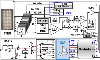
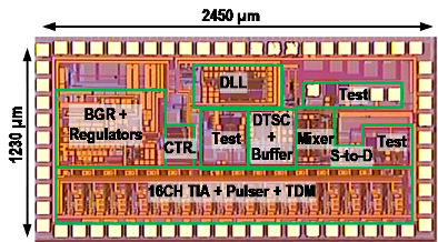
Real time cross-sectional image of the coronary arteries is needed for accurate monitoring of the lumen area during cardiovascular interventions, and intravascular ultrasound (IVUS) has become widely used for this purpose along with angiography and optical tomography. Current IVUS catheters, which have a diameter of 3.5 F, are not small enough to advance in narrow or severely occluded arteries, limiting the use of IVUS to only ~20% of cardiovascular interventions. Hence, miniaturization of the imaging devices on small catheters has been one of the major concerns in IVUS systems. We aim to reduce the size of the IVUS system so that it can eventually placed on a 0.014” guidewire, which is used in all minimally invasive cardiovascular interventions.
Single Chip Intracardiac Echocardiography (ICE) System with Cable Reduction
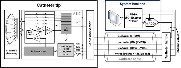
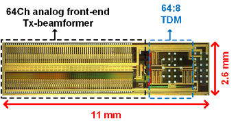
Intracardiac echocardiography (ICE) has become an important clinical modality in interventional ultrasound imaging. It requires a minimally invasive procedure with local anesthesia to place a catheter in the right side of the heart to guide interventions like valve repair, placement of stents, closure of atrial septal defects (ASD), and catheter- based ablation to treat atrial fibrillation. Current commercial ICE catheters offer a 2D or limited 3D field of view in spite of large number of interconnections, which are mainly determined by the number of array elements and ground connections. Reducing the number of connections by integrating electronics at the catheter tip would improve the overall system performance by capturing the receiver signals with higher fidelity, and transmitting them to the backend using higher quality cables. Cable reduction would also improve the mechanical flexibility of these single use catheters, and possibly lower the cost. Therefore, integrating electronics at the catheter tip would have a significant impact in the catheter-based ultrasound imaging applications.
Project funding: NIH
Associated students: Ahmad Rezvanitabar, Brian Chen
References
[1] J. Lim, C. Tekes, E. F. Arkan, A. Rezvanitabar, F. L. Degertekin and M. Ghovanloo, “Highly Integrated Guidewire Ultrasound Imaging System-on-a-Chip,” in IEEE Journal of Solid-State Circuits, vol. 55, no. 5, pp. 1310-1323, May 2020, doi: 10.1109/JSSC.2020.2967551.
[2] J. Lim, C. Tekes, F. L. Degertekin and M. Ghovanloo, “Towards a Reduced-Wire Interface for CMUT-Based Intravascular Ultrasound Imaging Systems,” in IEEE Transactions on Biomedical Circuits and Systems, vol. 11, no. 2, pp. 400-410, April 2017, doi: 10.1109/TBCAS.2016.2592525.
[3] J. Lim, C. Tekes, A. Rezvanitabar, E. F. Arkan, F. L. Degertekin and M. Ghovanloo, “Highly-integrated guidewire vascular ultrasound imaging system-on-a-chip,” 2018 IEEE Custom Integrated Circuits Conference (CICC), San Diego, CA, 2018, pp. 1-4, doi: 10.1109/CICC.2018.8357032.
[4] G. Jung et al., “A Reduced-Wire ICE Catheter ASIC With Tx Beamforming and Rx Time-Division Multiplexing,” in IEEE Transactions on Biomedical Circuits and Systems, vol. 12, no. 6, pp. 1246-1255, Dec. 2018, doi: 10.1109/TBCAS.2018.2881909.
[5] G. Jung et al., “Single-chip reduced-wire active catheter system with programmable transmit beamforming and receive time-division multiplexing for intracardiac echocardiography,” 2018 IEEE International Solid – State Circuits Conference – (ISSCC), San Francisco, CA, 2018, pp. 188-190, doi: 10.1109/ISSCC.2018.8310247.
[6] G. Jung et al., “Single-Chip Reduced-Wire CMUT-on-CMOS System for Intracardiac Echocardiography,” 2018 IEEE International Ultrasonics Symposium (IUS), Kobe, 2018, pp. 1-4, doi: 10.1109/ULTSYM.2018.8579915.