Force Integrated Readout and Active Tip for Atomic Force Microscopy
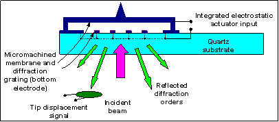
A membrane-based force sensor for probe microscopy with electrostatic actuation and integrated optical detection has recently been introduced by Degertekin group at Georgia Institute of Technology. This structure was originally developed as an opto-acoustic device for microphone and ultrasonic transducer applications. This new AFM structure is named as FIRAT for Force Sensing Integrated Read-out Active Tip. A schematic description of the FIRAT probe together with the incident laser beam, reflected diffraction orders and photodetectors is given in Figure 1.

Figure 1: Schematic description of the opto-acoustic pressure sensor with integrated actuation and interferometric optical readout that was introduced by Degertekin et al.
In order to create a new FIRAT probe, a platinum tip is built on a membrane (Figure 2) by using FIB.
First implementations of FIRAT showed that it can be used for direct measurement of interaction forces. Time domain interaction forces (TRIF) are directly measured as shown in Figure 3. Here, the FIRAT probe substrate oscillates as driven by a suitable actuator. Both the attractive and repulsive regions of the force curve are traced as the probe tip contacts the sample during each cycle.

Figure 3: The shape of the FIRAT probe during different phases of a cycle of the time resolved interaction force (TRIF) mode. The size and direction of the arrows indicate the speed and direction of motion of the FIRAT probe substrate. The substrate position and the FIRAT probe output signals are also shown.
Since the electrostatic forces act only on the probe membrane, the actuation speed as limited by the membrane dynamics is quite fast. Therefore, combined with array operation, the FIRAT probe will be useful for probe microscopy applications requiring high speeds. In this mode, the Z input of the piezo tube is disconnected and used only for x-y scan. The integrated electrostatic actuator is used for both oscillating the probe tip at 600 kHz and controlling the membrane bias level in order to keep the oscillation amplitude constant as the tapping mode images are formed.

Figure 4: Topography images of the 20 nm thick steps of the calibration grating at different imaging speeds measured by using the integrated electrostatic actuator of the FIRAT probe for vertical motion.
Since we are able to obtain the time-domain interaction force, we can get the sample properties by using a suitable model. Intermolecular distance, effective tip-sample Young’s Modulus, Poisson’s ratio and surface energy are important parameter for this relationship. The simulation results of our model and experimental results agree within 10% of error in contact duration as you can see in Figure 6.
People: Zehra Parlak, Rameen Hadizadeh, Kianoush Naeli, Giray Oral
Collaborators:
Funding: National Science Foundation


